
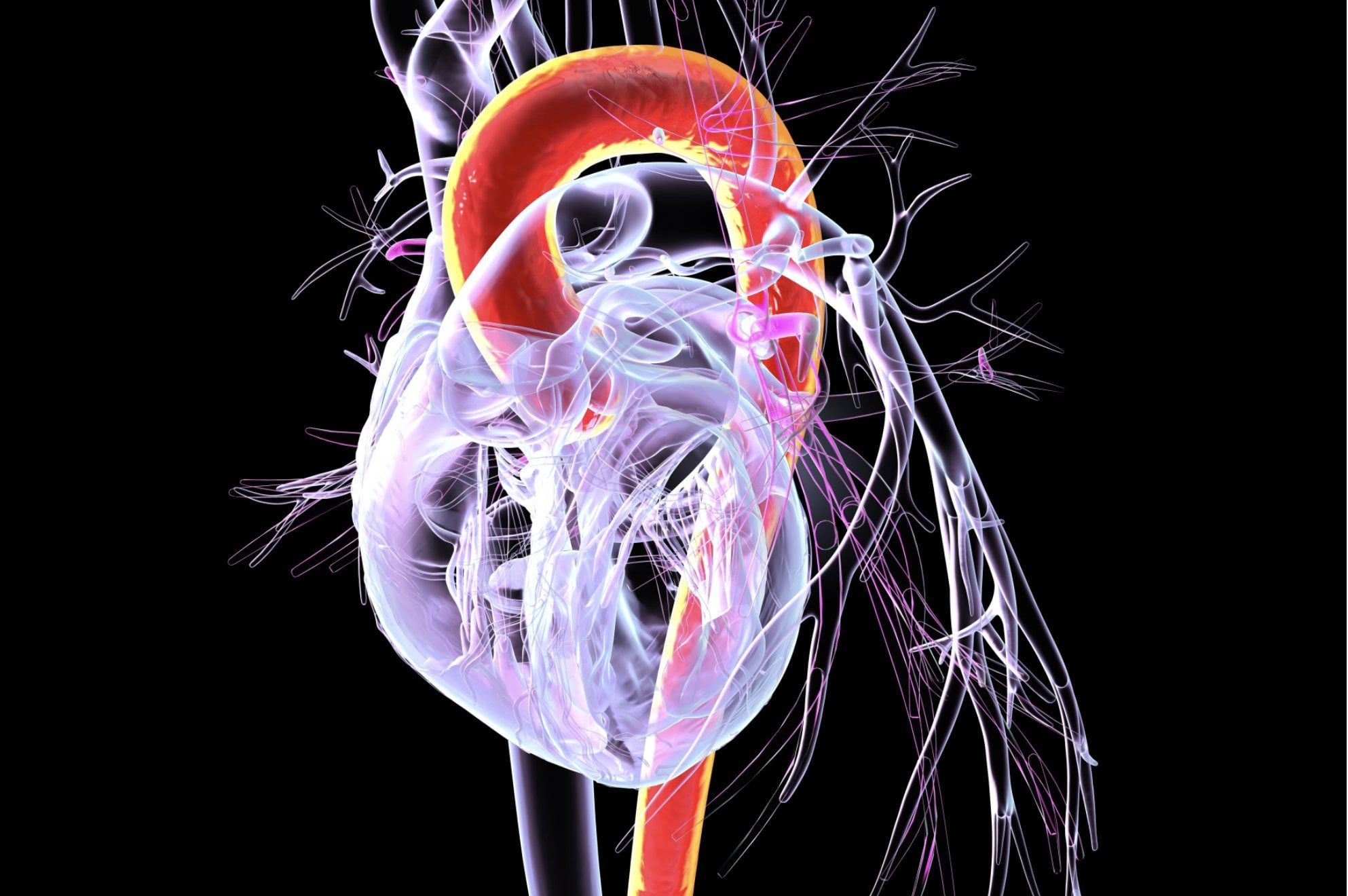
 A team at OSF HealthCare is developing an artificial intelligence-powered tool designed to improve how physicians measure and monitor the aorta, a breakthrough that could help doctors detect dangerous aneurysms earlier and make more informed decisions about when surgery is needed.
The project, led by pediatric cardiologist Mathew Bramlet, MD, at OSF HealthCare Children's Hospital of Illinois, combines expertise from OSF HealthCare, Jump Simulation and Education Center, the University of Illinois College of Medicine Peoria and University of Illinois engineering researchers. The technology creates a highly detailed three-dimensional "digital twin" of a patient's anatomy from MRI or CT scans, allowing software to automatically analyze the aorta in seconds instead of requiring lengthy manual measurements.
Get The Latest News!
Don't miss our top stories and need-to-know news everyday in your inbox.
The aortic arch project, funded through a Jump ARCHES grant, is aimed at improving longitudinal surveillance and the diagnosis and the tracking of aortic aneurysms, so they don’t tear or rupture. The aorta is the main and largest artery in the human body, carrying oxygen-rich blood from the left ventricle of the heart to the rest of the body. An aneurysm is a large bulge in that artery.
Accurate measurements are critical
Dr. Bramlet says measurements that document any expansion of the aneurysm need to be obtained with clear imaging with an expert eye that is generating reproducible measurements. Since high-risk surgery can hinge on one measurement, the accuracy of that measurement at one point in time, or over years as the growth of the aneurysm is monitored, becomes critically important. Among the complications doctors trying to prevent is an aortic dissection which recently killed Senator Lindsey Graham of North Carolina.
Currently, physicians often compare scans taken over many years to determine whether an aneurysm has grown enough to justify preventative surgery — a decision that carries significant risks if performed too early or too late. Manually measuring the aorta can take about 30 minutes and depends heavily on specialist expertise like that of Dr. Bramlet.
The new software automates that process, which Dr. Bramlet explains can generate up to 300 measurements in about 30 seconds while improving consistency.
“If we can capture this aorta and generate those same measurements with fancy new math and machine learning programs, we can distribute the expertise that I have accumulated over years and years and years, essentially into a program that would allow this type of analysis to be distributed at a larger scale.”
The automated approach also could allow researchers to analyze thousands of patient scans, helping develop more personalized treatment guidelines for conditions such as Marfan syndrome and Turner syndrome.
Aorta library will be created
The project is also creating a library of 3D digital models of healthy and diseased aortas that could eventually support automated diagnosis.
"The model library that we will have, where we can compare a diseased aorta to a normal aorta and look for the differences, it's a much easier automated process to get toward automated diagnosis, automated analysis and this is really the mindset of the entire lab,” says Dr. Bramlet.
Dr. Bramlet directs the Advanced Imaging and Modeling Lab at OSF Jump Simulation.
Beyond aortic disease, the underlying technology is designed to create digital models of multiple organs, opening the door to broader AI-assisted imaging analysis across medicine.
For Dr. Bramlet, it is exciting to see how this will be able to help provide better guidance for diagnosis and surgery determinations. The team is continuing to validate the technology through research while working toward clinical deployment – something Dr. Bramlet says is unique to OSF HealthCare.
“To have a hospital institution assign operational resources towards a clinical tool but I believe it is the commitment to serve people and to actually see the fruits of that labor being deployed within this institution really makes it a special place.”
In research conducted, this tool showed that automated 3D measurements closely match those done by clinicians, paving the way for a shift from traditional 2D imaging to more advanced 3D analysis.
Dr. Mathew Bramlet is also an associate professor of Clinical Pediatrics at the University of Illinois College of Medicine in Peoria.
